 |
| http://www.virtualmedicalcentre.com/uploads/VMC/DiseaseImages/2133_eye_anatomy_label_v2_700.jpg |
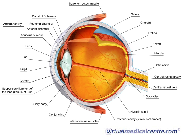
Cornea: front sixth clear layer in which all light must pass first when it enters the eye
Iris: colored part of the eye; an adjustable diaphragm around an opening called the pupil
Lens: clear, bi-convex structure which changes shape because it is attached to muscles in the ciliary body to fine-tune vision
Ciliary body: muscular area attached to the lends which contracts and relaxes to control the size of the lens for focusing
Optic nerve: conducts the electrical impulses from the light entering the eye to the brain
Choroid: second layer of the eye that contains the blood vessels that supply blood to structures of the eye
Sclera: tough, outermost layer of the eye which maintains the shape of the eye
Retina: innermost layer and light-sensing portion of the eye which contains rod cells (responsible for vision in low light) and cone cells (responsible for color vision and detail)
Macula: center of the retina which contains the fovea centralis (contains only cones and is responsible for seeing fine detail clearly
Myopia vs. hyperopia
Near-sightedness vs. far-sightedness
Myopia is a refractive defect of the eye in which light produces an image in front of the retina which translates to you are better at seeing objects close up than far away. Hyperopia is literally the exact opposite. Thus, the image is produced behind the retina which means that you can see objects better far way than close up. Myopia usually occurs with an eyeball that is longer or a cornea that is steeper than normal. Hyperopia usually occurs with an eyeball is shorter or the cornea has too little curvature.
LASIK surgery:
LASIK surgery is performed with a laser programmed to remove a defined amount of tissue from the cornea. The doctor will cut a flap on the top layer of the cornea which allows access to the deeper layers of the eye. They layer is used again to flatten certain tissue or to make the tissue steeper, depending on the needs of the patient. Finally, the flap is folded back into place and will heal on its own. This procedure corrects vision because it makes up for the discrepancies of the eye making sure the image will be focused on the retina, not ahead or behind it.